

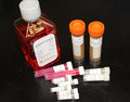


- Culture Primary Endothelial Cells M1168 is tested and optimized with mouse endothelial cell growth and proliferation. Use aseptic technique to prevent microbial contamination.
- Culture Primary Animal Cells All cell culture procedures must be conducted in a bio-safety cabinet.
- Freezing or Recovering Cells Cryo-preserved cells must be stored in liquid nitrogen or seeded immediately upon arrival. Coat sterile culture dishes or flasks with Gelatin-Based Coating Solution (Cell Biologics, Catalog No. 6950) for 2 min and then aspirate the excess solution before seeding cells.
- Handling Live Cultured Cells Keep a flask with 20 ml existing medium in 37°C CO2 incubator for 1 hour before replacing the medium. Either split the 90% confluent cells from a T25 flask to a T75 flask after 1 hour or let the cells grow in the T25 flask for 12-24 hours before split the cells to a T75 flask. Cells should be checked daily under a microscopy to verify appropriate cell morphology.
- Protocol for Flow Cytometry, Uptake of Dil-Ac-LDL and Tube Formation Assay and cell staining (Endothelial Cells) For best results, please carefully review the detailed protocol to use an unconjugated primary antibody. Incubate endothelial cells with Dil-Ac-LDL for 4 h at 37°C. Thaw reduced growth factor matrigel (BD: 354234) by submerging the bottle on ice and store the matrigel at 4 °C overnight.
- FAQ about Custom Endothelial Cell Isolation Primary endothelial cells produced from Cell Biologics display typical "cobblestone" or "spindle-shaped" morphology under light microscopy and show VE-cadherin (CD144) staining at cell-cell junction or Von Willebrand Factor staining in plasma. These cells also express VE-cadherin and CD31 as demonstrated by FACS analysis.
- Storage of Cell Biologics' Products Primary cells should never be kept at -20 °C freezer.
GFP Cell Preparation
· GFP-Labeled Primary Cells employing Amaxa nucleofector transfection methods are detached from the culture flask and immediately cryo-preserved in vials. Each vial contains 0.5-1.0x10⁶ cells per ml and is delivered frozen. GFP marked primary cells are verified by a fluorescent microscope.
· Cells can be seeded on 30-60 mm culture dishes ready for experiments (or cells may be expanded for 1-2 passages at a split ratio of 1:2) under the cell culture conditions specified by Cell Biologics. Repeated freezing and thawing of cells is not recommended.
Protocols for GFP-labeled Primary mammalian cells
· Harvest the cells by trypsinization. Count an aliquot of the trypsinized cells and determine cell density.
· Centrifuge the required number of cells (0.5-1.5 x10⁶ cells per sample) at 220xg for 5 minutes at room temperature.
· Resuspend the cell pellet carefully in 100 µl room temperature Nucleofector™ Solution per sample.
· Combine 100 µl of cell suspension with 5-10 µg pmaxGFP™ Vector (recommended for initial optimization).
· Transfer cell/DNA suspension into certified cuvette; sample must cover the bottom of the cuvette without air bubbles. Close the cuvette with the cap.
· Select the appropriate Nucleofector™ Program.
· Insert the cuvette with cell/DNA suspension into the Nucleofector™ Cuvette Holder and apply the selected program.
· Take the cuvette out of the holder once the program is finished.
· Add 500 µl of the pre-equilibrated culture media to the cuvette and gently transfer the sample immediately into the culture dish. Use the supplied pipettes and avoid repeated aspiration of the sample.
· Incubate the cells in a humidified 37°C/5% CO2 incubator until analysis. GFP expression is often detectable after only 4 – 8 hours but ideally, cells should be left undisturbed for 24 hours. GFP expression efficiency depends on different cell types.
Imported from the previous cellbiologics.com site.
